In vivo deep two-photon imaging of neural circuits with the fluorescent Ca2+ indicator Cal-590
05-Feb-2017
The Journal of Physiology, DOI: 10.1113/JP272790
The Journal of Physiology, online article
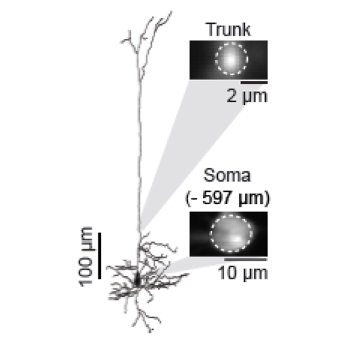
In vivo two-photon Ca2+ imaging has become an effective approach for the functional analysis of neuronal populations, individual neurons and subcellular neuronal compartments in the intact brain. When imaging individually labelled neurons, depth penetration can often reach up to 1 mm below the cortical surface. However, for densely labelled neuronal populations, imaging with single-cell resolution is largely restricted to the upper cortical layers in the mouse brain. Here, we review recent advances of deep two-photon Ca2+ imaging and the use of red-shifted fluorescent Ca2+ indicators as a promising strategy to increase the imaging depth, which takes advantage of reduced photon scattering at their long excitation and emission wavelengths. We describe results showing that the newly introduced fluorescent Ca2+-sensitive dye Cal-590 can be used to record in vivo neuronal activity in isolated cortical neurons and in neurons within populations in depths of up to 900 μm below the pial surface. Thus, the new approach allows the comprehensive functional mapping of all six cortical layers of the mouse brain. Specific features of Cal-590-based in vivo Ca2+ two-photon imaging include a good signal-to-noise ratio, fast kinetics and a linear dependence of the Ca2+ transients on the number of action potentials. Another area of application is dual-colour imaging by combining Cal-590 with other, shorter wavelength Ca2+ indicators such as OGB-1. Overall, Cal-590-based two-photon microscopy emerges as a promising tool for the recording of neuronal activity at depths that were previously inaccessible to functional imaging of neuronal circuits.


