Cellular, subcellular and functional in vivo labeling of the spinal cord using vital dyes
07-Feb-2013
Nature Protocols, 2013, doi:10.1038/nprot.2013.022, 8, 481–490, published on 07.02.2013
Nature Protocols, online article
Nature Protocols, online article
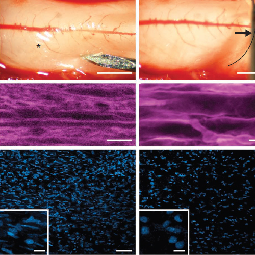
Here we provide a protocol for rapidly labeling different cell types, distinct subcellular compartments and key injury mediators in the spinal cord of living mice. This method is based on the application of synthetic vital dyes to the surgically exposed spinal cord. Suitable vital dyes applied in appropriate concentrations lead to reliable in vivo labeling, which can be combined with genetic tags and in many cases preserved for postfixation analysis. In combination with in vivo imaging, this approach allows the direct observation of central nervous system physiology and pathophysiology at the cellular, subcellular and functional level. Surgical exposure and preparation of the spinal cord can be achieved in less than 1 h, and then dyes need to be applied for 30–60 min before the labeled spinal cord can be imaged for several hours.


