A reversible form of axon damage in experimental autoimmune encephalomyelitis and multiple sclerosis
27-Mar-2011
Nature Medicine, 2011, doi:10.1038/nm.2324, 495–499 (2011) published on 27.03.2011
Nature Medicine, online article
Nature Medicine, online article
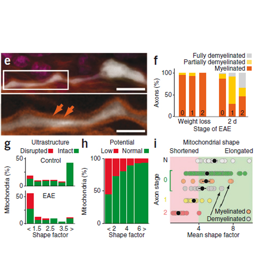
In multiple sclerosis, a common inflammatory disease of the central nervous system, immune-mediated axon damage is responsible for permanent neurological deficits1,2. How axon damage is initiated is not known. Here we use in vivo imaging to identify a previously undescribed variant of axon damage in a mouse model of multiple sclerosis. This process, termed ‘focal axonal degeneration’ (FAD), is characterized by sequential stages, beginning with focal swellings and progressing to axon fragmentation. Notably, most swollen axons persist unchanged for several days, and some recover spontaneously. Early stages of FAD can be observed in axons with intact myelin sheaths. Thus, contrary to the classical view2–6, demyelination—a hallmark of multiple sclerosis—is not a prerequisite for axon damage. Instead, focal intra-axonal mitochondrial pathology is the earliest ultrastructural sign of damage, and it precedes changes in axon morphology. Molecular imaging and pharmacological experiments show that macrophage-derived reactive oxygen and nitrogen species (ROS and RNS) can trigger mitochondrial pathology and initiate FAD. Indeed, neutralization of ROS and RNS rescues axons that have already entered the degenerative process. Finally, axonal changes consistent with FAD can be detected in acute human multiple sclerosis lesions. In summary, our data suggest that inflammatory axon damage might be spontaneously reversible and thus a potential target for therapy.


