Imaging of integrin alpha-v-beta-3 expression in patients with malignant glioma by [18F] Galacto-RGD positron emission tomography
01-Mar-2009
Neuro-Oncology, 2009, 11(6), 861-870, doi:10.1215/15228517-2009-024 published on 01.03.2009
Neuro-Oncology, online article
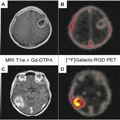
Inhibitors targeting the integrin {alpha}vβ3 are promising new agents currently tested in clinical trials for supplemental therapy of glioblastoma multiforme (GBM). The aim of our study was to evaluate 18F-labeled glycosylated Arg-Gly-Asp peptide ([18F]Galacto-RGD) PET for noninvasive imaging of {alpha}vβ3 expression in patients with GBM, suggesting eligibility for this kind of additional treatment. Patients with suspected or recurrent GBM were examined with [18F]Galacto-RGD PET. Standardized uptake values (SUVs) of tumor hotspots, galea, and blood pool were derived by region-of-interest analysis. [18F]Galacto-RGD PET images were fused with cranial MR images for image-guided surgery. Tumor samples taken from areas with intense tracer accumulation in the [18F]Galacto-RGD PET images and were analyzed histologically and immunohistochemically for {alpha}vβ3 integrin expression. While normal brain tissue did not show significant tracer accumulation (mean SUV, 0.09 ± 0.04), GBMs demonstrated significant but heterogeneous tracer uptake, with a maximum in the highly proliferating and infiltrating areas of tumors (mean SUV, 1.6 ± 0.5). Immunohistochemical staining was prominent in tumor microvessels as well as glial tumor cells. In areas of highly proliferating glial tumor cells, tracer uptake (SUVs) in the [18F]Galacto-RGD PET images correlated with immunohistochemical {alpha}vβ3 integrin expression of corresponding tumor samples. These data suggest that [18F] Galacto-RGD PET successfully identifies {alpha}vβ3 expression in patients with GBM and might be a promising tool for planning and monitoring individualized cancer therapies targeting this integrin.


